DNA
DNA (or deoxyribonucleic acid) is the means by which hereditary information is encoded within the cells of most living organisms. A small proportion of organisms encode their hereditary information using a related molecule called RNA (or ribonucleic acid), but for humans, our hereditary information is encoded in DNA.
DNA takes the form of a "double helix" (Lodish, et al., 2004, p. 103). This double helix is often thought of as a spiral structure with two side chains and rungs connecting them, similar to a spiral staircase (see Figure 2: DNA Structure). In reality, DNA is made up of repeating units called nucleotides. Each nucleotide is made up of three components: a phosphate group, a sugar molecule, and a base. The phosphate group is linked to the sugar molecule which is in turn is linked to the base. The phosphate group of each nucleotide is also bonded to the sugar molecule of the adjacent nucleotide forming a polynucleotide chain (Lodish, et al., 2004, p. 103).
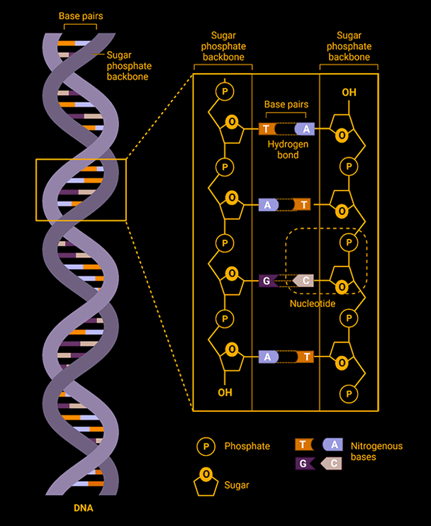
There are four bases which are commonly referred to by their first letter: A (adenine), T (thymine), G (guanine), and C (cytosine). The base is the important part of each nucleotide as it is the base that encodes the genetic information. The bases are sometimes referred to as "base pairs" as they exhibit what is known as Watson–Crick base pairing where A (adenine) always forms a base pairing with T (thymine) and G (guanine) always forms a base pairing with C (cytosine). This means that if there is an A (adenine) on one side of the DNA molecule there will always be a T (thymine) on the other side, and vice versa. Likewise, if there is a G (guanine) on one side of the DNA molecule there will always be a C (cytosine) on the other side, and vice versa (Mao, 2004). This base pairing is what allows DNA to replicate. The base pairs can detach from one another splitting the DNA molecule into two strands. The two strands can then rebuild their missing sides by assembling nucleotides with the complimentary bases to the existing bases (Lodish, et al., 2004, p. 133).
Types of DNA
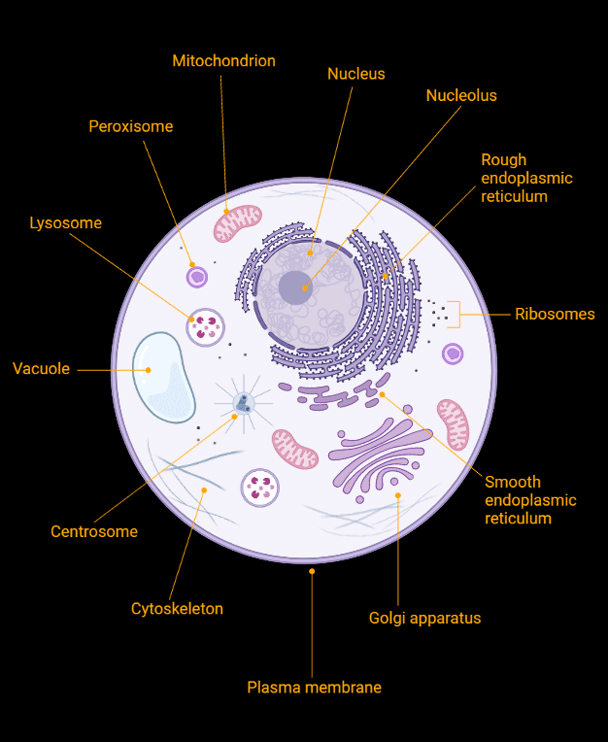
Our cells contain two different kinds of DNA: nuclear DNA (nDNA) and mitochondrial DNA (mtDNA). Nuclear DNA is comprised of DNA packaged into large X-shaped chromosomes and is contained in the nucleus of each of our cells. Mitochondrial DNA is packaged into small circular chromosomes contained in the mitochondria (singular: mitochondrion) which are present in the cytoplasm of each of our cells (see Figure 3: Cell Structure). Mitochondria are organelles (small organ-like structures) that are responsible for energy production within our cells (Bolohan, Ciorpac, Mățău, & Gorgan, 2016, p. 161).
Nuclear DNA (nDNA)
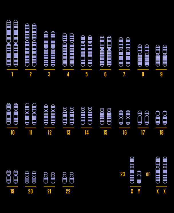
When we refer to a person's DNA we are usually referring to their nuclear DNA. It is a person's nuclear DNA that will determine how tall they are or what hair colour or eye colour they have. These traits are determined by genes. Genes come in different variants called alleles. Humans generally possess 23 pairs of chromosomes. 22 of these pairs are referred to as autosomal chromosomes and one pair is referred to as the sex chromosomes (see Figure 4: Human Karyotype). Humans inherit one of each pair of chromosomes from each of their parents, meaning that one of their two alleles came from their mother, and one came from their father (Bolohan, Ciorpac, Mățău, & Gorgan, 2016, p. 161).
We commonly think of DNA as being made up of genes, but genes actually only make up around 1% of our DNA (National Library of Medicine, 2021). Genes encode proteins. The remaining 99% of our DNA is made up of non-coding sequences. It was once thought that this non-coding DNA was "junk" and that it didn't have any function. It is now understood that much of this non-coding DNA performs important regulatory functions that determine when and where genes are turned on or off (National Library of Medicine, 2021).
Humans share 99.9% of their nuclear DNA (National Human Genome Research Institute, 2018). It is in the remaining 0.1% that a person's ancestry can be revealed.
Mitochondrial DNA (mtDNA)
Mitochondria possess DNA because prior to the evolution of multicellular life they existed as separate single-celled organisms. At some point in time a type of alphaproteobacteria entered the cell from which all plant, animal, and fungal life would evolve, a type of Asgard archaea, and began an endosymbiotic relationship (Zaremba-Niedzwiedzka, et al., 2017, p. 353). In this endosymbiotic relationship the host cell would supply the symbiont bacteria with glucose and in return the bacteria would supply the cell with its required energy source, Adenosine triphosphate (ATP) (Cavalier-Smith, 2006, p. 1943).
Except in the case of mitochondrial disease which affects energy production within cells, mitochondrial DNA does not determine a person's characteristics. However, mitochondrial DNA can be useful when researching your ancestry. Unlike nuclear DNA which is inherited from both parents, mitochondrial DNA is inherited solely from a person's mother (Bolohan, Ciorpac, Mățău, & Gorgan, 2016, p. 162).
Y-chromosomal DNA (Y-DNA)
Within the chromosomes of the nuclear DNA there are the sex chromosomes. Generally speaking, human females have two X-chromosomes and human males have one X-chromosome and one Y-chromosome. It is the SRY gene on the Y-chromosome that determines whether a person is male or female (National Library of Medicine, 2022). Just as mitochondrial DNA is passed from mother to child, the Y-chromosome is passed from father to son (Bolohan, Ciorpac, Mățău, & Gorgan, 2016, p. 162).
Note: There are cases where females do possess a Y-chromosome (National Library of Medicine, 2016) and cases where males do not due to the translocation of the SRY gene from the Y-chromosome to another chromosome (McElreavey & Cortes, 2001, p. 133).
Explore the Human Migration page next to see how nuclear, mitochondrial, and Y-chromosomal DNA can help you explore your ancestry.
Figure References:
Figure 2: DNA Structure - Original work created using BioRender.com
Figure 3: Cell Structure - Original work created using BioRender.com
Figure 4: Human Karyotype - Original work created using BioRender.com
References:
Bolohan, N., Ciorpac, M., Mățău, F., & Gorgan, D. L. (2016). Ancestral DNA - An incontestable source of data for Archaeology. Studia Antiqua et Archaeologica, 21(2), 157-188. Retrieved from Studia Antiqua et Archaeologica: http://saa.uaic.ro/
Cavalier-Smith, T. (2006). Origin of mitochondria by intracellular enslavement of a photosynthetic purple bacterium. Proc. R. Soc. B., 273(1596), 1943-1952. doi: https://doi.org/
Lodish, H., Berk, A., Matsudaira, P., Kaiser, C., Kreiger, M., Scott, M., Zipursky, L., & Darnell, J. (2004). Molecular Cell Biology, 5th Edition. New York: W. H. Freeman.
Mao, C. (2004). The Emergence of Complexity: Lessons from DNA. PLOS Biology, 2(12), e431. doi: https://doi.org/
Mathews, C. K., van Holde, K., Appling, D. R., & Anthony-Cahill, S. J. (2013). Biochemistry, 4th Edition. Toronto: Pearson Canada Inc.
McElreavey, K., & Cortes, L. S. (2001). X-Y translocations and sex differentiation. Seminars in Reproductive Medicine, 19(2), 133-140. doi: https://doi.org/
National Human Genome Research Institute. (2018, September 7). Genetics vs. Genomics Fact Sheet. Retrieved from National Human Genome Research Institute: https://www.genome.gov/
National Library of Medicine. (2016, November 1). Androgen insensitivity syndrome. Retrieved from Medline Plus: https://medlineplus.gov/
National Library of Medicine. (2021, January 19). What is noncoding DNA? Retrieved from MedlinePlus: https://medlineplus.gov/
National Library of Medicine. (2022, September 28). SRY gene. Retrieved from Medline Plus: https://medlineplus.gov/
Zaremba-Niedzwiedzka, K., Caceres, E. F., Saw, J. H., Bäckström, D., Juzokaite, L., Vancaester, E., Seitz, K. W., Anantharaman, K., Starnawski, P., Kjeldsen, K. U., Stott, M. B., Nunoura, T., Banfield, J. F., Schramm, A., Baker, B. J., Spang, A., & Ettema, T. J. G. (2017). Asgard archaea illuminate the origin of eukaryotic cellular complexity. Nature(541), 353-358. doi: https://doi.org/10.1038/